Science Milestone

THE HISTORY OF
ANTI-VEGF THERAPIES

Blood vessels grow everywhere in the human body, but aberrant growth can lead to diseases like cancer. Through painstaking research in the past few decades, scientists have figured out ways to control this vessel growth to help stop disease progression.

download pdf
Supported by

1930s - 1960s

Early clues linking blood vessel growth to tumors

In 1939, a group of scientists at Rochester University decided to investigate the ear of a rabbit. After transplanting a bit of carcinoma into the animal’s ear, they looked at the growth of blood vessels (angiogenesis) in the area. It turned out that the tumor growth was accompanied by the division of these blood vessels — and that if these vessels did not grow, the tumor wouldn’t grow either (1).

These findings, along with the quiet publication of a few others, pushed forward the idea that something blood vessel-related was important to the pathogenesis of cancer. In 1945, scientists at the National Cancer Institute counted the number of blood vessels that grew alongside transplanted tumors and found that blood vessel growth preceded tumor expansion (2).

As scientists became more obsessed with finding the molecules that governed these biological processes, people from different fields began to postulate how these blood vessels might grow. Some thought that a mysterious factor, possibly secreted from tumor cells, caused blood vessel growth. Others thought that the eye’s retina created something that similarly led to vessel growth, since blood vessels play a prominent role in the eye (3). In 1968, a group at Harvard Medical School showed that even sticking a filter in between transplanted cancer cells and the host wouldn’t stop blood vessels from growing at a frenetic pace, definitively proving that there was some mysterious traveling factor that caused angiogenesis to occur (4,5).

‍Professor Ernest Starling at the University College London coined the word “hormone” in 1905.  ‍Professor Ernest Starling at the University College London coined the word “hormone” in 1905.  
CREDIT: (Top Image) ISTOCK.COM/ DESIGN CELLS , (Bottom Image) ISTOCK.COM/ STELLA_E
(Top Image) Cancer cells release VEGF to promote the formation of new blood vessels that nourish and sustain tumor growth. (Bottom Image) Scientists discovered the link between blood vessel growth and tumor growth by looking in the ear of a rabbit.

1970s

A mysterious “factor”

The Aschheim-Zondek-Haus was home to a women’s clinic and a laboratory that developed the first laboratory pregnancy test in 1927.
CREDIT: CHILDREN’S HOSPITAL BOSTON
Judah Folkman was considered the “father of angiogenesis.”

With the emerging theory that cancers caused blood vessels to go haywire, the race was on to figure out exactly how. In 1971, a pediatric surgeon named Judah Folkman postulated that blocking blood vessel growth might stop cancer growth. While this theory was met with some uproar in the scientific community, Folkman’s lab simultaneously reported their initial efforts to isolate the mysterious compound responsible for blood vessel growth from different tumor models and cultured cells (6).

Folkman hypothesized that a diffusing factor would be plausible — this molecule would need to travel long distances in possibly turbulent blood vessels, after all. To identify what exactly this factor might be, Folkman’s lab homogenized a rat breast tumor and then tried to fractionate the homogenate through a gel. They then tested each fraction on whether it would stimulate blood vessel growth.

One fraction did indeed have strong angiogenic activity. The scientists dubbed the fraction “tumor angiogenesis factor” — a mixture of fats, proteins, and RNA. However, with the technology of the time, isolating a single protein or molecule from this fraction was extremely laborious.

1980s

Finding VEGF

In the years after Folkman’s group introduced the “tumor angiogenesis factor” to the scientific community, researchers continued to search for this mysterious factor that would stimulate blood vessel growth. Some research groups had isolated fibroblast growth factors (FGFs) that appeared to be potential candidates (7). However, FGFs weren’t secreted and seemed to stay firmly inside cells. As a result, the search continued and piqued the interest of Napoleone Ferrara, then a postdoctoral scholar at the University of California at San Francisco.

Ferrara was studying follicular stellate cells, which live in the pituitary gland. “Among the things that we found very intriguing was the fact these cells were in this close proximity to blood vessels,” he said. The team isolated the cells’ supernatant, essentially a soup containing all sorts of secreted molecules, and used a technique called high-performance liquid chromatography to separate the specific protein molecules in the supernatant (8).

After testing various protein fractions to see if they stimulated the growth of endothelial cells, which make up blood vessels, the team tried to microsequence the fraction to determine its amino acid sequence. They confirmed this fraction was comprised primarily of one unique protein. Then, they found that the protein had a previously unknown amino acid sequence, indicating that this was, indeed, a newly discovered protein. Since the protein only stimulated the growth of vascular endothelial cells, the scientists dubbed it “vascular endothelial growth factor,” or VEGF.

Ferrara, who had moved to Genentech, remembered finally getting the terminal amino acid sequence from a collaborator who surprised him at a dinner party. “I was stunned because I did not expect any of that,” he said. “I remember it as a great evening.”

‍The Hogben test indicated pregnancy if a female Xenopus frog laid eggs within 12 hours after injection with a women’s urine.  
CREDIT: PRINCE MAHIDOL AWARD
Napoleone Ferrara’s team discovered VEGF.

1990s

VEGF in all its forms

‍In 1977, Rosalyn Yalow was working at the Veterans Administration Hospital when she learned that she, along with two other American doctors, had been awarded the Nobel Prize in Physiology or Medicine for their discovery of the radioimmunoassay (19). ‍In 1977, Rosalyn Yalow was working at the Veterans Administration Hospital when she learned that she, along with two other American doctors, had been awarded the Nobel Prize in Physiology or Medicine for their discovery of the radioimmunoassay (19).
CREDIT: (Top Image)  ISTOCK.COM/ SVETAZI (Bottom Image) HARVARD OPHTHALMOLOGY
(Top Image) A high level of VEGF can encourage the growth of abnormal blood vessels underneath the retina, causing eye diseases such as macular degeneration. (Bottom Image) Joan Miller and her colleagues discovered how VEGF works in the eye.

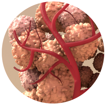
Isolating VEGF was merely one step. Next, Ferrara and his team wanted to see how important the factor was. To do this, the scientists knocked out the VEGF gene in mice. “It showed that even if you inactivated one of the two alleles, there is embryonic lethality,” said Ferrara. “This showed that this was a very important factor, not easily replaced.” Ferrara’s group, along with other scientists at the University of California at San Francisco, then isolated a receptor called FMS-like tyrosine kinase 1 that bound to VEGF with a very high affinity (9). As researchers continued to unravel VEGF biology, they isolated other members of the VEGF family, including VEGF-B, C, and D.

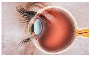
Meanwhile, scientists who studied the eye became very interested in how VEGF might fit into the progression of ocular diseases, some of which are caused by markedly increased blood vessel growth in the eyeball. One of them was Joan Miller, an ophthalmologist at Harvard Medical School who had just joined Folkman’s lab during her retina fellowship.

During her training, Miller developed nonhuman primate models of eye diseases. By using these models and studying retinal cells in a dish, Miller and the team found that increased levels of VEGF correlated with increased vascularization in the eye — and that, specifically, it was the retina that produced VEGF (10,11). Finally, the scientists showed that “you could take VEGF protein and inject it into the vitreous of normal animal eyes and develop new blood vessels,” Miller said. “So it was sufficient in and of itself to produce new blood vessels” (12).

Miller’s findings were recapitulated by a larger study showing that in human patients, VEGF was indeed associated with the growth of new blood vessels (13). With these results in hand, scientists began to test Folkman’s original 1971 theory — if blocking VEGF would in fact reduce tumor growth or, with the new knowledge that the molecule was heavily implicated in the eye and ocular diseases.

2000s

Blocking VEGF

‍Reproductive biologist Glenn Braunstein codeveloped the antibodies specific to hCG and its subunits at the National Institutes of Health.
CREDIT: ISTOCK.COM/ MARCIN KLAPCZYNSKI
Avastin is an anti-VEGF antibody developed by Genentech.

At Genentech, Ferrara’s interest in VEGF had persisted. First, his team engineered a monoclonal mouse antibody against VEGF, called an anti-VEGF antibody. Then, the team demonstrated that injecting this antibody into tumors would inhibit their growth. In particular, the more rapidly proliferating tumor, which was more dependent on angiogenesis, had a bigger decrease in growth. “It seemed to many improbable that blocking one factor would do much at all,” said Ferrara. “What we found was greatly exceeding what we expected.”

Ferrara shared the anti-VEGF antibody with Miller and her team, who injected it into the eyes of the animal models. They found that the antibody completely inhibited the growth of new blood vessels in the eye (14). “It was almost like one of those things where you pinch yourself because we didn’t think it would be that effective,” said Miller.

This anti-VEGF antibody became Genentech’s Avastin, a medication approved in the US in 2004 for treating colon cancer. Other technologies that similarly blocked VEGF, such as anti-VEGF antibody fragments, sprouted in biotechnology labs around the world.

2012-present

Anti-VEGF therapies in the clinic

‍Reproductive biologist Glenn Braunstein codeveloped the antibodies specific to hCG and its subunits at the National Institutes of Health.
CREDIT: ISTOCK.COM/ ARTUR PLAWGO
Scientists are interested in developing new therapies such as gene therapies for angiogenesis-related diseases.

Avastin was soon approved for the treatment of several other cancers, including those of the lung, ovaries, and kidney. Another VEGF blocker, an aptamer — a small strand of nucleic acids that tightly binds to a target — called Macugen, was developed by OSI Pharmaceuticals and approved by the FDA to treat macular degeneration in 2004. Then, in 2006, an anti-VEGF antibody fragment named Lucentis was developed by Genentech and approved again for macular degeneration.

While these therapies provided new hope for patients, they had their drawbacks. The macular degeneration therapies, for example, required monthly eye injections. In addition, blocking VEGF even locally had side effects, like high blood pressure or an increased risk of bleeding, though perhaps not as catastrophic as what people had thought.

Ferrara, Miller, and others are continuing their research on VEGF, anti-VEGF, and the biology associated with blood vessel formation. Miller noted that gene therapies, which are often one and done, might be a nice alternative for ocular diseases where the goal is to reduce the number of injections needed for vessel formation blockage. Ferrara, whose research interests have pivoted to also focus more on eye diseases, is studying how VEGF inhibitors can be engineered to last longer in the eye.

For Ferrara, the road ahead does not diminish the work already done — even with the newer therapeutic technologies like immunotherapies or gene therapies that are becoming available. “It is really rewarding that twenty years later, this drug [anti-VEGF] is not obsolete,” he said.

REFERENCES 

  1. G, I. A. Vascularization of the brown-pearce rabbit epithelioma transplant as seen in the transparent ear chamber. Am J Roentg 42, 891 (1939).
  2. Algire, G. H., Chalkley, H. W., Legallais, F. Y. & Park, H. D. Vasculae Reactions of Normal and Malignant Tissues in Vivo. I. Vascular Reactions of Mice to Wounds and to Normal and Neoplastic Transplants. JNCI J. Natl. Cancer Inst. 6, 73–85 (1945).
  3. Michaelson, I. The mode of development of the vascular system of the retina, with some observations on its significance for certain retinal diseases. in (1948). at <https://www.semanticscholar.org/paper/The-mode-of-development-of-the-vascular-system-of-Michaelson/69f11a4f0ce22a430e 1c8483d437c529f1e6c287>
  4. Ehrmann, R. L. & Knoth, M. Choriocarcinoma. Transfilter stimulation of vasoproliferation in the hamster cheek pouch. Studied by light and electron microscopy. J. Natl. Cancer Inst. 41, 1329–1341 (1968).
  5. Greenblatt, M. & Shubi, P. Tumor angiogenesis: transfilter diffusion studies in the hamster by the transparent chamber technique. J. Natl. Cancer Inst. 41, 111–124 (1968).
  6. Folkman, J., Merler, E., Abernathy, C. & Williams, G. Isolation of a tumor factor responsible for angiogenesis. J. Exp. Med. 133, 275–288 (1971).
  7. Ferrara, N. VEGF and the quest for tumour angiogenesis factors. Nat. Rev. Cancer 2, 795–803 (2002).
  8. Ferrara, N. & Henzel, W. J. Pituitary follicular cells secrete a novel heparin-binding growth factor specific for vascular endothelial cells. Biochem. Biophys. Res. Commun. 161, 851–858 (1989).
  9. de Vries, C. et al. The fms-like tyrosine kinase, a receptor for vascular endothelial growth factor. Science 255, 989–991 (1992).
  10. Miller, J. W. et al. Vascular Endothelial Growth Factor/Vascular Permeability Factor Is Temporally and Spatially Correlated with Ocular Angiogenesis in a Primate Model. Am. J. Pathol. 145, 574–584 (1994).
  11. Shima, D. T. et al. Cloning and mRNA expression of vascular endothelial growth factor in ischemic retinas of Macaca fascicularis. Invest. Ophthalmol. Vis. Sci. 37, 1334–1340 (1996).
  12. Tolentino, M. J. et al. Vascular endothelial growth factor is sufficient to produce iris neovascularization and neovascular glaucoma in a nonhuman primate. Arch. Ophthalmol. Chic. Ill 1960 114, 964–970 (1996).
  13. Aiello, L. P. et al. Vascular endothelial growth factor in ocular fluid of patients with diabetic retinopathy and other retinal disorders. N. Engl. J. Med. 331, 1480–1487 (1994).
  14. Adamis, A. P. et al. Inhibition of vascular endothelial growth factor prevents retinal ischemia-associated iris neovascularization in a nonhuman primate. Arch. Ophthalmol. Chic. Ill 1960 114, 66–71 (1996).